GI Endoscopy

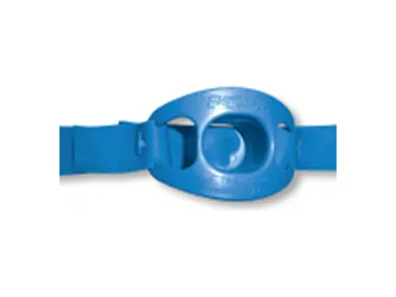
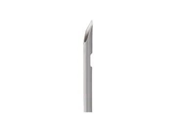
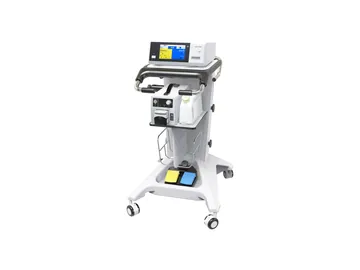
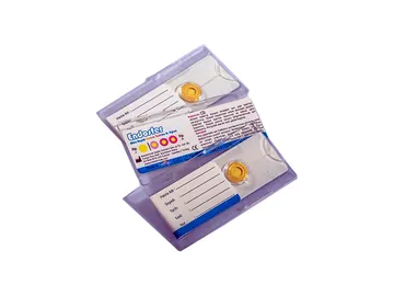
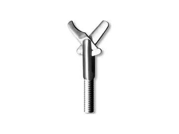
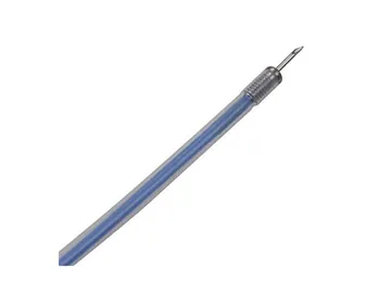
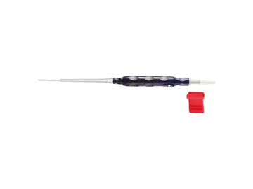
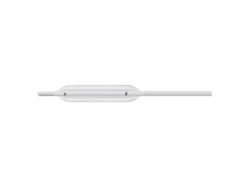
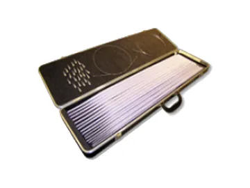
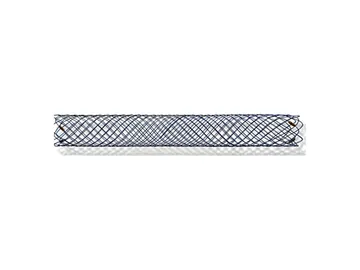
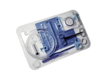
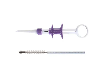
You find the items of your trolley in eight groups: polypectomy, hemostasis, dilation, stents. In the polypectomy and hemostasis diagrams, each product appears at the step where it is used.

This page is intended for healthcare professionals.
GI Endoscopy
Procedure

Colonic polypectomy and EMR, step by step

A small polyp and a large flat lesion in the colon: six steps from assessment to clip closure.

haustralumenfold
Schematic longitudinal section, not to scale. Lumen and lesion sizes are proportional; the layers of the lower wall are drawn thick so they can be read.
  1. 01

    Lesion assessment and classification

    Size and Paris morphology are assessed; if deep submucosal invasion is suspected, en bloc resection or surgery is planned instead of standard polypectomy.

    Clinical detail

    The lesion is examined closely: size, Paris morphology (pedunculated 0-Ip, sessile 0-Is, flat 0-IIa), surface and vessel pattern, site and access. If findings suggest deep submucosal invasion, en bloc resection or surgery is planned instead of standard polypectomy. The scope is positioned so that the lesion lies in line with the working channel, usually at 5 to 6 o'clock on the image; a biopsy is taken when needed.

  2. 02

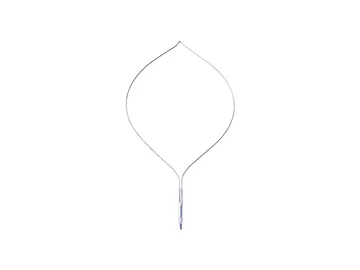
    Small polyp: cold snare

    A polyp up to 9 mm is cut without current; ESGE 2024 recommends cold snare, not cold forceps, given incomplete resection rates.

    ESGE 2024

    Clinical detail

    A polyp up to 9 mm is captured with a 1 to 2 mm rim of normal mucosa and cut by closing the snare without current. ESGE 2024 recommends cold snare polypectomy at this size and recommends against cold biopsy forceps excision because of its high rate of incomplete resection. The resected polyp is usually suctioned into a polyp trap.

    Source: Ferlitsch M et al. ESGE, Endoscopy 2024;56:516-545 (PMID 38670139)

  3. 03

    Lifting injection

    Fluid lifts the submucosa, separating the lesion from the muscle layer; ESGE recommends adding dye; a lesion that does not lift suggests fibrosis or invasion.

    Clinical detail

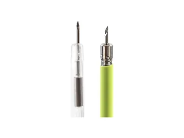
    For a large or flat lesion the needle enters the submucosa through the mucosa at an oblique angle; the fluid expands the submucosa and forms a cushion that separates the lesion from the muscle layer. ESGE recommends adding a dye to the fluid, which shows the extent of the cushion, the lesion margins and any deep mural injury. A lesion that does not lift suggests fibrosis or invasion and may change the plan.

  4. 04

    Hot snare resection (EMR)

    The lesion is cut with current; ESGE 2024 recommends hot snare for non-pedunculated adenomas of 10 to 19 mm and conventional EMR from 20 mm.

    ESGE 2024

    Clinical detail

    The lifted lesion is captured, the snare is closed and the tissue is cut with electrosurgical current; lesions up to about 20 mm are usually removed en bloc, larger ones piecemeal. ESGE 2024 recommends hot snare for non-pedunculated adenomas of 10 to 19 mm and conventional EMR from 20 mm; after piecemeal EMR the margins are treated with snare-tip soft coagulation. For a pedunculated polyp with a head of 20 mm or a stalk of 10 mm or more, the stalk is first treated with dilute adrenaline or a mechanical method.

  5. 05

    Polyp retrieval

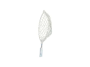
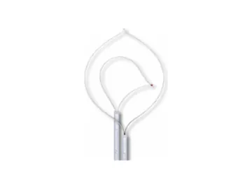
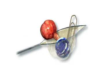
    Tissue is retrieved for histopathology: large pieces in a retrieval net, small ones in a trap; a net snare holds the polyp in one pass.

    Clinical detail

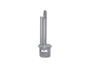
    Resected tissue is retrieved for histopathology. A large piece is held in a through-the-channel retrieval net and brought out without withdrawing the scope; small pieces are collected in a multi-chamber trap on the suction line. A net snare holds the polyp in its net in the same pass.

  6. 06

    Defect check and clip closure

    ESGE 2024 recommends clip closure of the defect after EMR of lesions of 20 mm or more in the right colon to reduce delayed bleeding.

    ESGE 2024 Pohl 2019

    Clinical detail

    The defect is inspected for residual tissue and muscularis propria injury (target sign); deep mural injury is closed with clips. ESGE 2024 recommends prophylactic clip closure of the defect after EMR of lesions of 20 mm or more in the right colon to reduce delayed bleeding. Intraprocedural bleeding is controlled with snare-tip coagulation or clips, combined with dilute adrenaline injection when needed.

    Source: Pohl H et al. Gastroenterology 2019;157:977-984 (PMID 30885778)

This section is intended for healthcare professionals. The steps are summarised from ESGE (2017, 2024) and US Multi-Society Task Force (2020) recommendations; indication, technique and product choice are the physician's decision, and each product's instructions for use apply.
Procedure

Endoscopic haemostasis in non-variceal upper GI bleeding, step by step

A bleeding peptic ulcer in the stomach: five steps from finding the source to the check and the H. pylori test.

rugaelumen actively bleeding ulcervisible vessel
Schematic section of the gastric antrum, not to scale. Ulcer, vessel and wall layers are enlarged so they can be read; blood is drawn in a dark, plain tone.
  1. 01

    Finding the source and Forrest classification

    Endoscopy follows haemodynamic resuscitation within 24 hours; the ulcer is graded by Forrest, and Ia, Ib and IIa lesions receive endoscopic therapy.

    ESGE 2026

    Clinical detail

    Endoscopy is performed within 24 hours after haemodynamic resuscitation; ESGE 2026 advises against endoscopy within 12 hours unless the patient stays unstable despite resuscitation. The stomach and duodenum are washed and inspected and the ulcer is graded by Forrest: spurting (Ia) or oozing (Ib) bleeding and a non-bleeding visible vessel (IIa) receive endoscopic therapy; an adherent clot (IIb) is removed to see what lies beneath, while a flat pigmented spot (IIc) and a clean base (III) need no therapy. If the source is variceal the pathway changes: ESGE recommends band ligation for oesophageal varices.

    Source: Gralnek IM et al. ESGE 2026, Endoscopy 2026;58:899-924 (PMID 42127996)

  2. 02

    Injection: slowing the bleeding

    Dilute epinephrine slows bleeding only temporarily; ESGE advises against epinephrine alone in active bleeding, and a second mechanical or contact thermal method follows.

    ESGE 2021

    Clinical detail

    Dilute epinephrine (adrenaline) is injected in small amounts around the bleeding vessel and into the ulcer base; the vessel constricts, the swollen tissue presses on it, bleeding slows and the view clears. The effect is temporary: ESGE advises against epinephrine alone in active bleeding, and a second mechanical or contact thermal method follows. For a non-bleeding visible vessel, sclerosant injection alone is among the options. ESGE 2026 does not list fibrin glue among initial treatment methods.

    Source: Gralnek IM et al. ESGE 2021, Endoscopy 2021;53:300-332 (PMID 33567467)

  3. 03

    Mechanical haemostasis: clips

    The clip grips the vessel from both sides; ESGE 2026 suggests the over-the-scope (OTS) clip as an alternative initial monotherapy for Ia and Ib ulcers.

    ESGE 2026

    Clinical detail

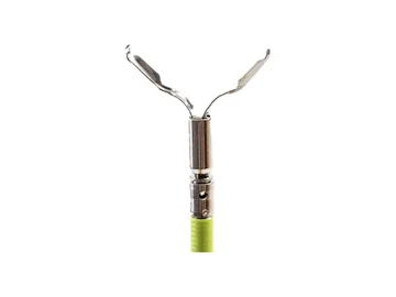
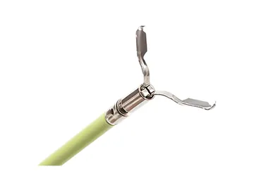
    The clip is advanced through the working channel, opened, pressed perpendicular onto the ulcer base over the visible vessel, closed so that it grips the vessel from both sides, and released; more clips are added when needed. A reopenable clip can be opened and closed until it sits right. On a hard, fibrotic base or at a tangential angle a clip may not hold; thermal therapy or an over-the-scope (OTS) clip is then considered. ESGE 2026 suggests the OTS clip as an alternative initial monotherapy for Ia and Ib ulcers.

  4. 04

    Thermal haemostasis: haemostatic forceps

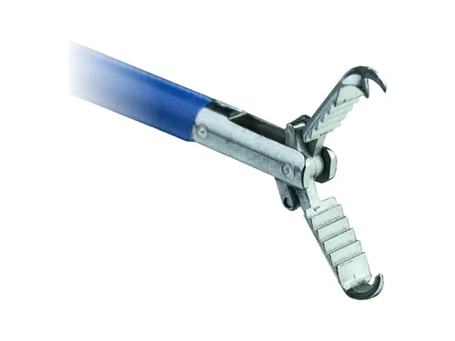
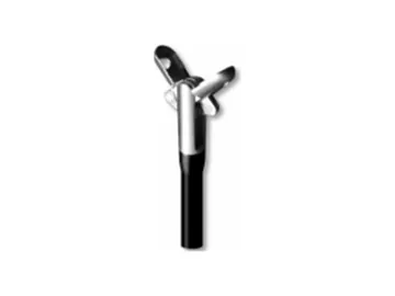
    The haemostatic forceps grasps the vessel and applies soft coagulation; ESGE 2026 states it may be used as monotherapy for Ia, Ib and IIa ulcers.

    ESGE 2026 Toka 2019

    Clinical detail

    The haemostatic forceps grasps the visible vessel gently, lifts it slightly off the ulcer base and soft coagulation is applied; haemostasis is considered achieved when the vessel is flattened and no longer visible. ESGE 2026 states that haemostatic forceps with soft coagulation may be used as monotherapy for Ia, Ib and IIa ulcers; AGA mentions it as an option in difficult locations and on a fibrotic base. In a randomised trial with monopolar forceps, initial haemostasis was higher than with clips.

    Source: Toka B et al. Gastrointest Endosc 2019;89:792-802 (PMID 30342026)

  5. 05

    Check, H. pylori and aftercare

    High-dose PPI is given for 72 hours after haemostasis; H. pylori is tested for in the acute setting and the test is repeated if negative.

    Gisbert 2006

    Clinical detail

    The ulcer is washed to confirm that bleeding has stopped. ESGE recommends testing for Helicobacter pylori in the acute setting and repeating the test if it is negative; biopsy-based tests lose sensitivity during bleeding. Biopsies come from the antrum and corpus, not from the ulcer. After haemostasis high-dose PPI is given for 72 hours and routine second-look endoscopy is not performed. Rebleeding calls for repeat endoscopy and an OTS clip; if that fails, transcatheter embolisation, and surgery where it is not available.

    Source: Gisbert JP, Abraira V. Am J Gastroenterol 2006;101:848-863 (PMID 16494583)

This section is intended for healthcare professionals. The steps are summarised from ESGE (2021, 2026), ACG (2021) and AGA (2020) recommendations; variceal bleeding follows a separate pathway and is not covered here. Indication, agent, technique and product choice are the physician's decision, and each product's instructions for use apply.
GI Endoscopy

Support in this category

Taught physician to physician, supported in the room for the first cases, supplied nationwide.