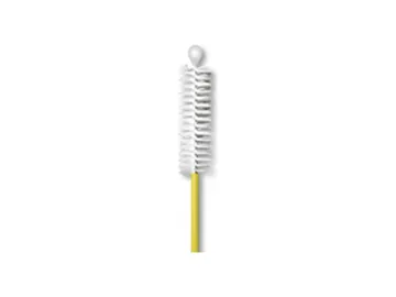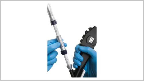
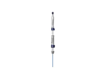
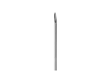
Bronchoscopy & EBUS

EBUS TBNA and TBNB needles for mediastinal staging; a conventional TBNA needle is coming soon. The needle selector matches the type a guideline recommends for a target with the needles we supply.

This page is intended for healthcare professionals.

Bronchoscopy & EBUS
Procedure

An EBUS-TBNA staging procedure, step by step

EBUS-TBNA for mediastinal staging in suspected lung cancer: six steps from the systematic survey in the airway to preparing the sample, and the conventional path used when EBUS is not available.

right lungleft lungtumourtracheacarinaaortapulmonary arterypulmonary arterySVC 2R2L4R4L10R10L11R11L7
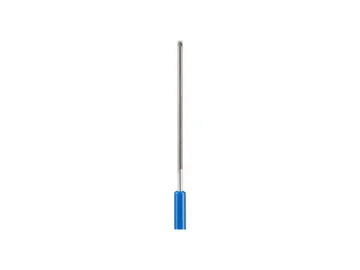
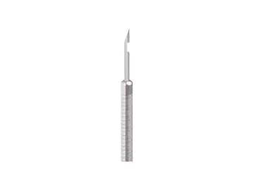
Schematic front view, not to scale (patient's right on the left): trachea, main bronchi, great vessels and IASLC lymph node stations; tumour in the right upper lobe. Bottom right: convex probe B-mode image of the subcarinal (7) station; bottom left: product close-up.
  1. 01

    Airway and systematic survey

    Stations are surveyed on the IASLC map in the order N3, N2, N1; ERS/ESGE/ESTS 2026 suggests systematic rather than targeted staging.

    ERS/ESGE 2026

    Clinical detail

    The convex probe EBUS bronchoscope is passed through the mouth into the trachea; its tip transducer is placed against the tracheal or bronchial wall, and the tip balloon is filled with water when needed. The stations are surveyed in a systematic order on the IASLC map: N3 (contralateral), then N2 (ipsilateral mediastinal), then N1 (hilar and interlobar). For a right upper lobe tumour the order may be 4L, then 7 and 4R, then 10R or 11R; when the same needle is used, this order aims to prevent cells being carried between stations and upstaging the patient. ERS/ESGE/ESTS 2026 suggests systematic rather than targeted staging and, where possible, EBUS combined with EUS(-B) sampling from the oesophagus.

    Source: Korevaar DA et al. ERS/ESGE/ESTS, Eur Respir J 2026;68(2) (PMID 42167778)

  2. 02

    Assessing the node, checking the path with Doppler

    With all four independent signs of metastasis absent, 96% of nodes were not metastatic; imaging does not replace sampling; Doppler checks the needle path.

    Fujiwara 2010

    Clinical detail

    The subcarinal (7) node is imaged from the medial wall of the right or left main bronchus and its short axis is measured. Round shape, distinct margin, heterogeneous echogenicity and the coagulation necrosis sign were reported as independent predictors of metastasis; when all four were absent, 96% of nodes were not metastatic. The image helps choose the node to sample; it does not replace sampling. The needle path is checked for vessels with colour Doppler; the right pulmonary artery and the left atrium lie around the subcarinal region.

    Source: Fujiwara T et al. Chest 2010;138:641-647 (PMID 20382710)

  3. 03

    Placing the needle and puncture

    The needle advances into the node between cartilage rings; per the instructions for use the stylet is pulled back at most 5 mm while advancing.

    Chest 2016

    Clinical detail

    The needle sheath is clicked onto the working channel of the bronchoscope and locked; the sheath is advanced until its tip shows on the endoscopic image, then fixed. The needle is advanced into the node between the cartilage rings, followed as a bright line on the ultrasound image. Per the instructions for use, the stylet is pulled back by no more than 5 mm while advancing and removed completely once the tip is inside the node. ERS/ESGE/ESTS 2026 considers 21G and 22G TBNA needles the standard and finds insufficient evidence for the structural use of other sizes and needle types.

    Source: Wahidi MM et al. CHEST, Chest 2016;149:816-835 (PMID 26402427)

  4. 04

    Sampling: stylet, suction, to-and-fro

    With the stylet out, the needle moves to and fro in the node; randomised trials found no difference between suction and no suction (physician's choice).

    Casal 2012 Scholten 2017

    Clinical detail

    With the stylet out, the needle is moved to and fro inside the node; the strokes can be directed to different parts of the node. Suction is the physician's choice: negative pressure with the VacLok syringe, or no suction (capillary technique). Randomised trials found no difference in adequacy or diagnosis between samples taken with and without suction, nor between sampling with and without the stylet. If suction is on, the syringe stopcock is closed before the needle leaves the node.

    Source: Casal RF et al. Chest 2012;142:568-573 (PMID 22156610); Scholten EL et al. Chest 2017;151:636-642 (PMID 27769775)

  5. 05

    Sample: ROSE and preparation

    The aspirate goes onto slides, the rest into a cell block or formalin; in a randomised trial ROSE reduced punctures at similar diagnostic sensitivity.

    Oki 2013 Gilbert 2025

    Clinical detail

    The needle is drawn into the sheath, locked and removed from the bronchoscope; the aspirate is expressed onto slides with the syringe or by reinserting the stylet, and the rest is flushed with saline into a cell block or formalin tube. Rapid on-site evaluation (ROSE) tells adequacy during the procedure; in a randomised trial ROSE reduced the number of punctures and the need for additional procedures, with similar diagnostic sensitivity. If molecular testing is planned, how the material is divided is agreed with pathology beforehand; CHEST 2024 gives separate recommendations on acquisition and handling.

    Source: Oki M et al. Respiration 2013;85:486-492 (PMID 23571718); Gilbert CR et al. Chest 2025;167:899-909 (PMID 39343294)

  6. 06

    Another pass, the next station, more tissue

    For tissue architecture, a TBNB needle or cryobiopsy may be considered; ERS/ESGE/ESTS 2026 finds cryobiopsy evidence still insufficient; the product label does not mention it.

    Lee 2008 Zhang 2021

    Clinical detail

    In a study without ROSE, sensitivity for malignant stations reached 95% at the third aspiration and the fourth added nothing. Once the sample is adequate, the next station follows. Where tissue architecture is needed, as in lymphoma, sarcoidosis or rare tumours, a TBNB needle that takes a core or mediastinal cryobiopsy may be considered; in a randomised trial cryobiopsy raised the diagnostic yield in rare tumours and benign disease. ERS/ESGE/ESTS 2026 still finds the evidence insufficient for the structural use of cryobiopsy; the decision is the physician's.

    Source: Lee HS et al. Chest 2008;134:368-374 (PMID 18263688); Zhang J et al. Eur Respir J 2021;58(6) (PMID 33958432)

    Cryobiopsy track: in a 90-patient prospective series, the 22G SonoTip TopGain® EBUS TBNB was used in 45 patients to open a track in the node, and a 1.1 mm cryoprobe followed it (Oruqaj G et al. ERJ Open Res 2026, PMID 42111651). The product label does not mention cryobiopsy; the cryoprobe is a separate device.

  7. 07

    Without EBUS: conventional TBNA

    Without an EBUS bronchoscope, large central nodes are sampled on CT landmarks without an ultrasound image; the entry point is planned on the CT map.

    Clinical detail

    Without an EBUS bronchoscope, large central nodes such as the subcarinal node are sampled on CT landmarks without an ultrasound image: the needle passes through a chosen point on the carina or the bronchial wall, between cartilage rings, and the sample is taken by aspiration. Because the target and the vessels cannot be followed on an image, the entry point is planned beforehand on the CT map.

    AConventional TBNA needle (coming soon)

This section is intended for healthcare professionals. The steps are summarised from the ERS/ESGE/ESTS (2026) and CHEST (2016, 2024) guidelines and the products' instructions for use; indication, technique and product choice are the physician's decision, and each product's instructions for use apply.

Needle selector

TBNA or TBNB? EBUS needles by target

First the difference between the two needle types, then the type the guidelines recommend for each target, and last our needles of that type. The guideline rows name the needle type; our needles are matched only by purpose and tip.

Two needle types, two different samples

The tip geometry decides what comes back. The drawings are schematic.

The guideline in brief. For diagnosing and staging lung cancer, ERS/ESGE/ESTS 2026 regards 21G or 22G TBNA needles as standard and finds the evidence insufficient for the structured use of other needle types and of cryobiopsy [1].

Other guideline statements

When malignancy is suspected, CHEST 2024 suggests 21G or 22G over 19G, at least four passes and ROSE [2]. In lymphoma and sarcoidosis, diagnostic yield was higher with core needles (TBNB), but the evidence is at meta-analysis level and of low certainty [3] [4].

Choose your target

Each row shows the needle type the guideline recommends for that target and the level of evidence. When you choose a row, our needles of that type are brought forward below.

Target
Cryobiopsy has no row here: ERS/ESGE/ESTS 2026 finds the evidence insufficient for its structured use.
How to read the level of evidence (GRADE)
Strong recommendation
What the guideline panel words as "recommend": most informed physicians would choose this for most patients.
Conditional (weak) recommendation
"Suggest": the choice may vary with the patient, the centre and the resources.
High quality
High confidence that the true effect lies close to the estimate; further research is unlikely to change it.
Moderate quality
The true effect is probably close to the estimate, but may be substantially different.
Low quality
Confidence in the estimate is limited; the true effect may be substantially different.
Very low quality
Very little confidence in the estimate; usually based on observational or indirect data.

Guidelines grade a recommendation on two axes: the strength of the recommendation and the quality of the evidence behind it. A strong recommendation can rest on low quality evidence, and a conditional one on high quality evidence. [6]

Our needles of this type

Matching uses only the purpose and tip recorded for each product; it is not a recommendation.

3 orderable needle families

Coming soon

  • Conventional TBNA Needle TBNAUsed with a bronchoscope without ultrasound guidance. 20, 21 and 22G; 15° bevelled tip, side hole; 100 or 120 cm. No catalogue number yet.

Write to us for information

All dimensions and comparison 7 models
Dimensions of all models
ModelProductPurposeTipShaftScopeStyletUltrasound visibilitySUT
GUS-45-18-022EBUS ProTBNAFacet CutStainless steelOlympus BF-UC190F and BF-UC180F, FujifilmRound, nitinolNot statedGHC1790
GUS-41-18-022EBUS ProTBNAFacet CutStainless steelPentaxRound, nitinolNot statedGHC1790
GUS-42-18-022ProFlex EBUSTBNAFacet CutNitinolPentaxRound, nitinolNot statedGHC1790
GUB-45-18-022TopGain EBUSTBNBCrown cut, 3 pointStainless steelOlympus BF-UC190F and BF-UC180F, FujifilmRound, nitinolLaser engravingGHC1790
GUB-46-18-022TopGain EBUSTBNBCrown cut, 3 pointNitinolOlympus BF-UC190F and BF-UC180F, FujifilmRound, nitinolLaser engravingNo code
GUB-42-18-022TopGain EBUSTBNBCrown cut, 3 pointNitinolPentaxRound, nitinolLaser engravingNo code
(coming soon)Conventional TBNATBNA, without ultrasound15° bevel, side holeMetal or Teflon sheathBronchoscopeNot statedNot statedNot stated

Dimensions from the manufacturer's catalogues and instructions for use registered in Türkiye; SUT (Turkish reimbursement) codes from the national device registry export (03.10.2026). "Not stated" means our documents do not give the value. The guideline rows are at needle-type level and are not a recommendation for any product listed. The instructions for use decide selection and compatibility; needle choice is the physician's decision. This section is intended for healthcare professionals.

References (6)
  1. Guideline (GRADE) Korevaar DA et al. ERS/ESGE/ESTS guidelines on endobronchial and oesophageal endosonography for the diagnosis and staging of lung cancer. Eur Respir J 2026;68(2). PMID 42167778. doi.org/10.1183/13993003.00097-2026
  2. Guideline (systematic review) Gilbert CR et al. Acquisition and handling of EBUS transbronchial needle samples: CHEST guideline. Chest 2024;167:899-909. PMID 39343294. doi.org/10.1016/j.chest.2024.08.056
  3. Meta-analysis, 11 studies, 1,434 patients Li SP et al. Transbronchial needle core biopsy vs aspiration under EBUS guidance: a meta-analysis. Eur Respir Rev 2026;35(180). PMID 42270118. doi.org/10.1183/16000617.0001-2026
  4. Meta-analysis, 6 studies, 646 patients Gupta N et al. Franseen tip vs non-Franseen tip needle in EBUS transbronchial needle sampling: systematic review and meta-analysis. J Bronchology Interv Pulmonol 2025. PMID 41216678. doi.org/10.1097/LBR.0000000000001032
  5. Multicentre RCT, 154 patients Kramer T et al. EBUS by 22G TBNB versus regular 22G TBNA needles: a randomized clinical trial. Lung Cancer 2023;185:107362. PMID 37757575. doi.org/10.1016/j.lungcan.2023.107362
  6. Methods paper Guyatt GH et al. GRADE: an emerging consensus on rating quality of evidence and strength of recommendations. BMJ 2008;336:924-926. PMID 18436948. doi.org/10.1136/bmj.39489.470347.AD

Bronchoscopy & EBUS

Support in this category

Taught physician to physician, supported in the room for the first cases, supplied nationwide.