ERCP
You find the items of an ERCP case in eight steps, from cannulation to drainage. Each product appears in the diagram at the step where it is used.
This page is intended for healthcare professionals.
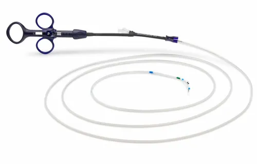
You find the items of an ERCP case in eight steps, from cannulation to drainage. Each product appears in the diagram at the step where it is used.
This page is intended for healthcare professionals.
Eight steps in a typical common bile duct stone case, from reaching the papilla to drainage.
The side-viewing duodenoscope reaches the duodenum and is withdrawn to the short position, facing the papilla; a rectal NSAID is given unless contraindicated.
The patient usually lies prone or semi-prone; sedation or anaesthesia is planned in advance and, without contraindication, a rectal NSAID is given just before the procedure. The side-viewing duodenoscope passes the stomach and pylorus into the second part of the duodenum, is rotated clockwise and withdrawn into the short position, and the papilla is brought face on.
The guidewire is advanced through the common bile duct; ESGE recommends the wire-guided technique for primary cannulation because it lowers post-ERCP pancreatitis risk.
The sphincterotome approaches the papilla along the bile duct axis, at 11 to 12 o'clock on the endoscopic image, and the guidewire is advanced through the common bile duct into the intrahepatic ducts. ESGE recommends the wire-guided technique for primary cannulation because it lowers the risk of post-ERCP pancreatitis.
ACannulation: sphincterotome or catheter
With the wire in place, contrast is injected at low pressure; a stone shows under fluoroscopy as a filling defect.
With the wire in place, contrast is injected at low pressure; under fluoroscopy the common bile duct, hepatic ducts, cystic duct and gallbladder fill. A stone shows as a filling defect the contrast cannot fill. Contrast given while a balloon occludes the duct is an occlusion cholangiogram; in a wide duct a small stone can be missed.
Source: Itoi T et al. Dig Endosc 2010;22 Suppl 1:S85-89 (PMID 20590779)
The papilla is opened in short, stepwise cuts between 11 and 1 o'clock; ESGE recommends blended rather than pure cutting current.
The cutting wire is gently tensioned and the papilla is opened in short, stepwise cuts within the safe zone between 11 and 1 o'clock; ESGE recommends blended rather than pure cutting current. For a difficult stone ESGE recommends adding papillary large-balloon dilation of 12 mm or more to a limited sphincterotomy, without exceeding the distal duct diameter.
Source: Manes G et al. ESGE, Endoscopy 2019;51:472-491 (PMID 30943551)
A balloon sweeps the duct and a basket catches stones it cannot hold; a final sweep or occlusion cholangiogram confirms a clear duct.
A balloon inflated above the stone sweeps the duct towards the papilla; a stone the balloon cannot hold is caught in a basket. A final balloon sweep or an occlusion cholangiogram shows that the duct is clear.
A stone that will not pass whole is crushed in a basket; ESGE recommends this when sphincterotomy and large-balloon dilation fail or are unsuitable.
A stone that will not pass the papilla whole is caught in a basket and crushed against a metal sheath; the fragments are removed with a balloon or basket. ESGE recommends it when sphincterotomy and large-balloon dilation fail or are unsuitable.
An indeterminate stricture is sampled with a cytology brush and a benign one can be balloon-dilated; without a stricture this step is skipped.
An indeterminate stricture is sampled with a cytology brush over the wire; a benign stricture can be dilated with a balloon over the wire. In a stone case without a stricture this step is skipped.
ACytology brush
BDilation balloon
A plastic stent is placed for an uncleared duct or stricture; at high pancreatitis risk ESGE recommends a prophylactic pancreatic stent plus rectal NSAID.
If the duct is not cleared or a stricture is present, a plastic stent is placed; a metal stent for a malignant stricture or a nasobiliary catheter for temporary external drainage are the alternatives. At high risk of pancreatitis ESGE recommends a prophylactic pancreatic stent in addition to a rectal NSAID; this is the physician's decision.
Source: Dumonceau JM et al. ESGE, Endoscopy 2020;52:127-149 (PMID 31863440)
APlastic stent
Also at this step
BMetal stent (coming soon)
CPancreatic stent
DNasobiliary catheter
This section is intended for healthcare professionals. The steps are summarised from ESGE and ASGE guidelines; indication, technique and product choice are the physician's decision, and each product's instructions for use apply.
Stainless steel and Nitinol families, with straight or angled tips in 0.025 and 0.035 inch.
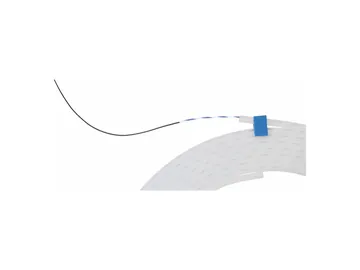
A range of single-use guidewires used to maintain and direct access across biliary and pancreatic strictures during ERCP, in stainless steel and in Nitinol.

A single-use guidewire for selective cannulation of the biliary ducts and for catheter introduction and exchange during endoscopic biliary procedures.
A version for Billroth II anatomy, short-wire series, and a series with a preloaded guidewire.
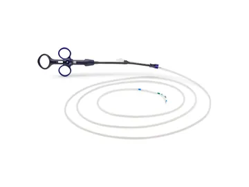
A single-use sphincterotome with a rotatable catheter tip for endoscopic sphincterotomy of the biliary or pancreatic sphincter.
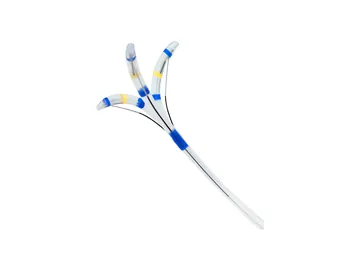
A single-use sphincterotome indicated for cannulation of the biliary ducts and transendoscopic sphincterotomy of the papilla of Vater and/or the sphincter of Oddi.
One balloon that reaches three diameters, single-size and three-stage models, and a short-wire series.
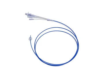
A single-use multi-stage extraction balloon used to sweep stones and debris from the bile duct after sphincterotomy.
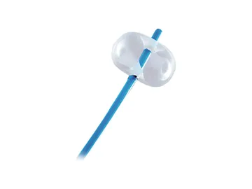
A single-use balloon catheter for the endoscopic removal of biliary stones, supplied sterile.
Braided or monofilament wires, braided baskets suit mechanical lithotripsy, and Nitinol and stainless steel baskets come on a short-wire system.
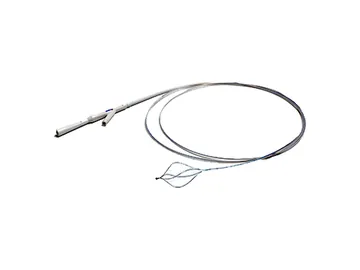
A single-use wire basket used to capture and remove bile duct stones, in braided and monofilament versions.
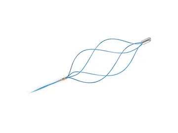
A single-use basket for the endoscopic removal of biliary stones and foreign bodies, in Nitinol and stainless steel.
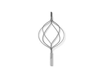
A mechanical lithotripsy device used to fragment large common bile duct stones that cannot be removed intact during ERCP.
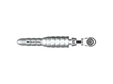
A lithotripsy system for large or impacted common bile duct stones: a reusable handle with single-use basket and spiral components, for planned and emergency cases.
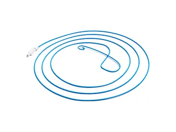
A nasobiliary drainage catheter (ENBD) placed during ERCP for temporary external drainage of the bile duct through the nasal passage.
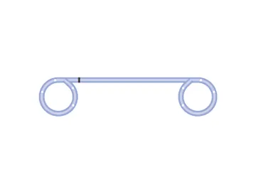
Single-use plastic drainage stents for the bile duct and for EUS-guided pseudocyst drainage, including double-pigtail designs.
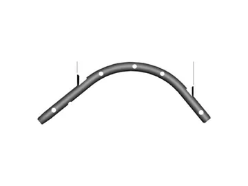
Single-use pancreatic stents in PTFE for drainage of the pancreatic duct, supplied with a positioning sleeve.
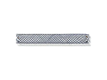
Single-wire-design self-expanding metal stents (SEMS) made of Nitinol for relieving malignant strictures in the biliary tree.
Coming Soon