Coming Soon Endoscopic Ultrasound · AI-System
mAI Companion®
A second pair of eyes in pancreatic EUS: two AI models on the live image. The decision stays with you.
This page is intended for healthcare professionals.
A second pair of eyes in pancreatic EUS: two AI models on the live image. The decision stays with you.
This page is intended for healthcare professionals.
EUS images the pancreas from the wall of the stomach and duodenum, and the result depends on the examiner: staying oriented, covering head, body and tail completely and noticing a small lesion in real time take practice. mAI Companion addresses these two points, coverage and detection. The examiner stays in charge.
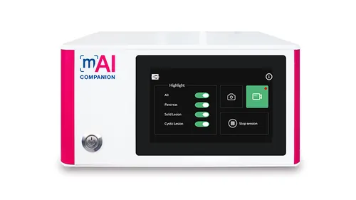
mAI Companion takes the video signal from your EUS processor and analyses it in real time. Its two models:

Recognises head, body and tail of the pancreas in real time, guides the coverage of each region and shows at a glance how long each region has been observed.

Marks solid and cystic areas that may be lesions the moment they appear on screen; you decide what to sample.
The software is hardware-independent and runs on an additional screen with a small form factor, so the existing EUS setup and workflow stay as they are. Images and video clips can be recorded and used in the report. The analysis runs locally, without sending data outside, and the device has carried the CE mark under the EU MDR since February 2026.
mAI Companion in action (animation by Medi-Globe)
The image count is Medi-Globe's own figure. The reader study was published in Endoscopy in August 2026 and tested a pre-commercial version of the software on recorded videos.
Annotated images from hundreds of real EUS procedures; the manufacturer states that the system was validated on these images and developed with endoscopists.
See the source → 70.8% → 77.1%57 endosonographers each read 38 EUS video clips with and without AI assistance of a pre-commercial version of the software; diagnostic accuracy for solid pancreatic tumours rose from 70.8% to 77.1%, mainly in clips with small and briefly visible lesions.
See the source →Sources: Endoscopy 2026, Montanelli, 57 endosonographers
Updated: 30.09.2026